Brodmann was right. Structure and function inform each other.
I saw this post, with this quote: “High-resolution activity maps of PFC did NOT align with cytoarchitecturally defined subregions.” Is this a profound, provocative finding that provides strong evidence for the irrelevance of cytoarchitecture?
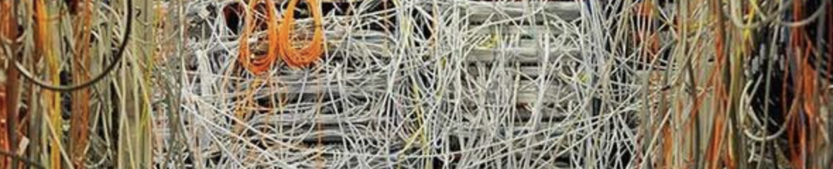
I’m a big fan of high quality neuroanatomy. Korbinian Brodmann’s work gave us a framework and a set of landmarks for cortical organization. Careful neuroanatomists have elaborated upon that, making corrections along the way, and provided important details for understanding brain function, all underpinned with the understanding that structure and function inform each other. The best neuroanatomists reveal order in the brain, where it previously looked like spaghetti wiring.
My favorite neuroanatomist is Andreas Burkhalter. He showed us organization like this (ref 1, ref 2):
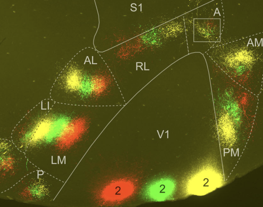
His work is tightly linked with function. Measuring visual responses in vivo, making precision tracer injections, studying synaptic properties in vitro, and detailed analysis.

So when a paper claims that activity maps are not aligned with cytoarchitecture, I wonder what they actually did and what they found. Are they upsetting a central premise? Or are they making a more specific observation?
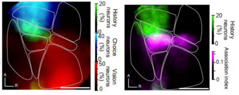
In the paper that kicked off the thread, the authors used a series of Neuropixels recordings in headfixed behaving mice, and analyzed the spontaneous spiking activity and task-related activity. They assigned units recorded to specific areas based the dye-labeled electrode tracks. Specifically, they estimated where they were on a common reference atlas using software. So they didn’t actually do cytoarchitectural analysis in the study to delineate areas. Other people in the thread cited some papers looking at the question which did nice cytoarchitectural work in individual animals. But that wasn’t done here.
I think the atlas approach is useful, but when trying to make specific claims about cytoarchitecture, especially precise boundaries between adjacent areas, the lack of precision can be a problem. Brains vary in size and shape. The paper on the software they used actually discussed this, quite well in fact, so I’ll just paste it here:
Functional landmarks in individual animals can be a better way to precisely identify cortical areas. Common landmarks, like skull landmarks, are okay for some applications, but not very precise. Especially for very small cortical areas < 0.5 mm across. For example, we provided this data in Riichiro’s recent eLife paper (this specific data and analysis was done by coauthor L. Townsend). Aligning to functional landmarks reveals highly regular mouse-to-mouse organization.
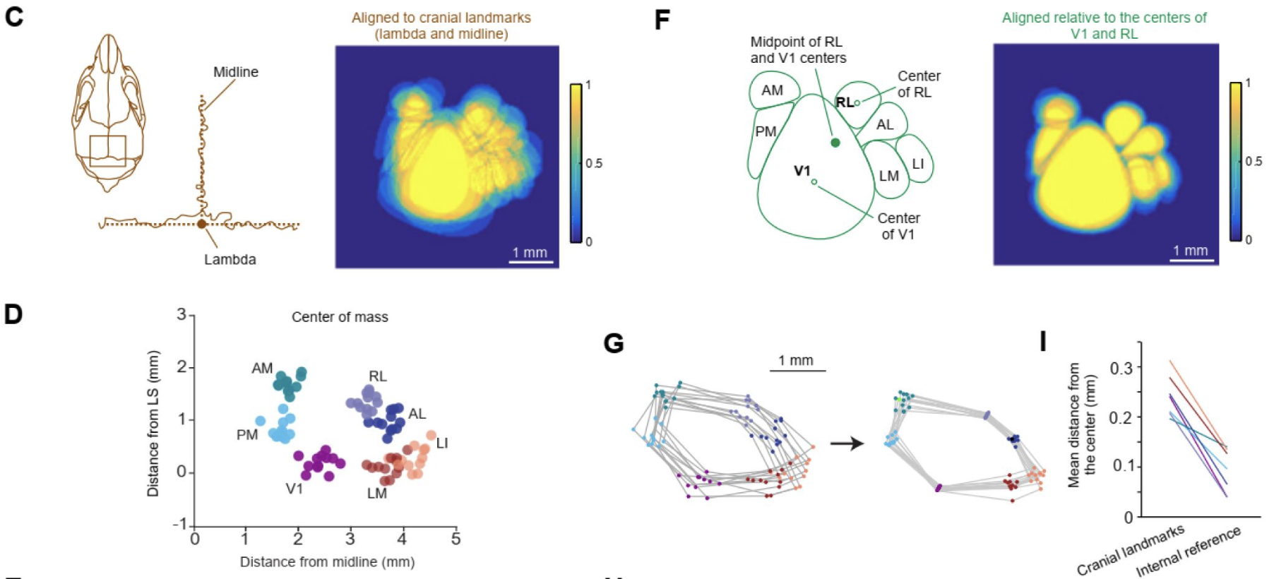
When you map things carefully, in individual animals, you can be very precise, and functional area borders can be quite striking. This is another figure from Riichiro’s paper (scale bar is 1 mm):

The rigor of the assignment of units to specific areas is one concern, another is the biases in extracellular recording and spike sorting. These biases can yield a dataset with relatively high spike rates, from similar neuron types, and these biases can influence the activity-based analysis, making the revealed relationships look more robust due to undersampling of more heterogeneous units.
Ultimately I disagree with the authors’ claim that “these findings challenge the traditional emphasis on cytoarchitecture, instead pointing to intrinsic connectivity as a primary organizing principle.” This is a false dichotomy. Of course connectivity matters, and the anatomists have taught us a tremendous amount about connectivity. The paper presents interesting analysis, with their self-organizing map (SOM) approach. There is definitely value in the paper and the work that the authors did. However, the paper does not provide strong evidence for the irrelevance of cytoarchitecture.