Scattering in tissue
Post by Jeffrey Stirman
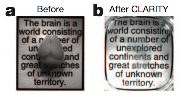
The opacity of the brain is one barrier to optically imaging individual neurons and their connections. Scattering in tissue is the main reason tissue is not transparent; absorption also plays a role but much less so. Perfusing tissue with a substance to match the index of refraction throughout the preparation (and thus decrease scattering) is one approach, and although index matching isn’t a new strategy, just getting rid of the membranes is. The most recent method to achieving tissue transparency (Chung et al., 2013), takes this approach to great effect.
A nice paper discussing tissue transparency is Johnsen and Widder, 1999. Scattering in tissue is dominated by Mie scattering which is the scattering of light by particles of a size on the same order as the wavelength of light (Rayleigh scattering is for particles much smaller than the wavelength): cells, nuclei, and organelles all fit in this category. Furthermore, the lipid membranes encasing these structures have a significantly different refractive index (~1.5) than the surrounding medium. It is this change in refractive index of these particles that lead to scattering. Simply, as the difference in refractive index between the surrounding medium and the object increases, so too does the scattering. The relationship with wavelength can be complicated and range from about lambda^-4 to lambda^0.2 (lambda = the wavelength of light used) depending on the size of the particle, but overall the higher the wavelength, the less scattering (one of the benefits of 2-photon imaging).
A couple of nice papers from Mourant et al. (1998 & 2000) discuss and explore in more detail the dominant scattering centers in tissue. They found that at small angles, most of the scattering was dominated by the nucleus and at larger angles the smaller structures such as mitochondria. One conclusion from all this is perhaps it might not be sufficient to homogenize the refractive index of the tissue if those lipid membranes still exist (as earlier attempts had done). In fact, the best way to achieve tissue clarity for imaging is to remove the objects that cause the scattering. This is exactly what Kwanghun Chung did! By first crosslinking most of the proteins, DNA, and other biological entities (not the lipids), then cross-linking them all in a hydrogel structure, he was able to use a detergent extraction process (electric field assisted) to remove the lipid membranes and thereby removing the cause of most of the scattering centers. Since multiple rounds of antibody staining can be performed on the cleared tissue, this process seems to have achieved clarity while preserving most of the interesting biology.


some irony that the Scale papers got flack for not referencing older work on clearing tissue, whereas this one gets all the hype… even managing to get past nature’s general standard of not publishing methods papers.