Field of view = 3.5 mm with cellular resolution
Posted in Hardware

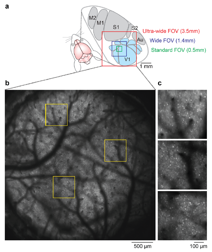
In this preprint, Stirman et al. report achieving a 3.5 mm field of view with 2-photon excitation with cellular resolution. The design involves custom scan optics and a custom objective built in the SLAB. It’s an air immersion objective with about a 9.5 mm working distance. Rapid dissemination is a priority. If you’re interested, contact slab (unc.edu).
Interesting paper … for the multiplexing part of the paper, have you looked at the pre-amplified time traces on the oscilloscope (maybe in averaging mode) to see how they look like and how fast they decay?
Yes, we have. They look like the depiction in Fig 3 here:
http://www.becker-hickl.com/pdf/ampmt.pdf
What’s the acquisition framerate and resolution in the 3.5mm FOV case? Are they the same as in the 1.4 mm case?
Do you think that this resolution is sufficient to do sophisticated whole-visual cortex analyses, or are we thinking more of cellular-resolution intrinsic-imaging-type experiments (outside of the mode-2 subfields)?
Finally, I’m surprised that you were able to express GCamp6s via viral injection on that huge surface. Any trick to it?
The data for the 3.5mm FOV above is from a transgenic mouse, not a viral injection.
As you can imagine, since the 3.5 mm FOV is ~150-fold more area than a more common 250×250 micron FOV ((pi*(1.75^2))/(0.25^2)=154), something has to give. That’s why we designed the system with temporally multiplexed, independently positionable imaging beams to image subregions (and/or different depths) at conventional resolutions and framerates.
That said, we’ve done some scanning of the 3.5mm FOV with a single beam, with a few pixels per cell, at < 1 frame/s (we have to zoom in a bit to get to 1 frame/s), and still detect single cell calcium transients with good S:N (similar to the 1.4 mm FOV data shown in the preprint). This is with normal raster scanning. For scanning the full FOV at higher temporal resolution, some sort of optimized scan path could help. To cover more cortical area at higher frame rates, using both beams makes sense. For example, two 1.5 mm FOVs centered in different portions of cortex (which can overlap) can cover a lot of cortex. Thanks for asking about this. We'll do some more characterization of this mode and add it to the manuscript.
…speaking of transgenic mice.
Are you willing to elaborate on that? 😉
No problem. The mice are at jax now.
[…] field of view isn’t going to be 3.5 mm wide, of course. That takes more extensive customization. But for conventional systems, this objective […]
This preprint has been updated now. Lots of new information including quad scanning and arbitrary line scanning.