Collection optics: How big is big enough?
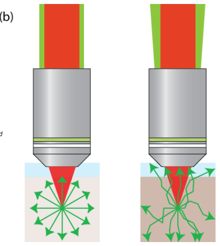
To advance deep imaging in multiphoton microscopy, it is essential to attempt to collect each photon generated in the scattering tissue. Large-diameter collection optics (50 mm or larger) have been suggested for efficient collection, as the emission of the signals at the back aperture of the objective could diverge greatly due to the scattering.
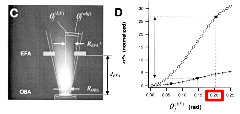
In 2001, Oheim et al. showed that the divergent angle of the emission (referred to as effective field angle, called Θƒ(EF) in the paper) of the then-new Olympus XLMPlanFluor 20× objective could be larger than 0.2 rad (11.5 deg). That measurement was used to rationalize the use of large-diameter collection optics.
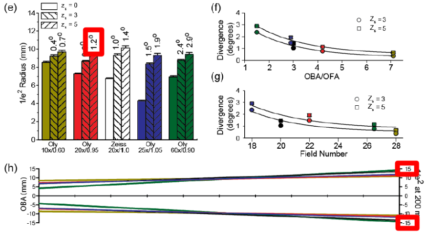
However, a different view was presented in 2015. Singh et al. showed that the broadening of the emission divergence is minimal: 0.021 rad (1.2 deg) for the same objective that Oheim et al. tested. This measurement was used to arguing against the necessity of the over-sized collection optics. Singh et al. noted that “we also find that the divergence of the fluorescence beam exiting the objective back aperture (OBA) is much less than previously reported. In general, 25 or 30 mm optics are sufficient for the detection pathway, provided they are situated relatively close to the objective”. The discrepancy between these groups could be due to the usage of different samples, which possibly gave rise to very different scattering properties (Oheim et al. used brain tissue, Singh et al. used a tissue phantom with polystyrene microspheres).
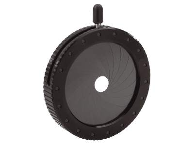
The question is to use large collection optics or not? The collection efficiency of two-photon microscopy depends not only on the diameter of the collection optics, but also the objective and the optical properties of the tissue. Labs use different combinations of objectives and samples, thus effective optimization strategies are unlikely to be uniform across labs. A simple procedure to test the significance of the divergence angle is to set up an adjustable iris diaphragm (e.g., one of these) in the collection pathway.
By measuring your signal as a function of the openness of the iris, you can get a better sense of what size of the collection optics is sufficient for your system.
(Post by Che-Hang Yu)
Zinter & Levene 2011 (10.1364/OE.19.015348) have simulated photon trajectories for the Olympus 20x 0.95 in Zemax. In Fig 5b they plot Collected Fluorescence vs Collection Angle, and it seems that fluorescence doesn’t increase beyond 10-11 degrees, which is pretty similar to the Oheim et al measurements.
>The discrepancy between these groups could be due to the usage of different samples, which possibly gave rise to very different scattering properties (Oheim et al. used brain tissue, Singh et al. used a tissue phantom with polystyrene microspheres).
Specifically they used 1 um microspheres at 800nm illumination, or 1.25 lambda scatterers. These will produce a moderate g value (anisotropy of scattering). Under those conditions, collection favors photons with fewer scattering events, which means photons that stay close to the optical axis and therefore have low angles out the back aperture of the objective. Reported g values for brain vary widely (0.85 to > 0.95), but at longer wavelengths g gets large and most deeper photons go through multiple scattering events before collection.
During my PhD studies, I looked at time of flight for backscattered photons in media with 1.25 lambda and 12.25 lambda scatterers using OCT [1]. I was surprised to find that for smaller scatters, most multiply scattered photons still had path lengths that were near the ballistic (unscattered) path length, even when they were on average scattered several times. When we looked at larger scatterers, the higher anisotropy (less deflection per scattering event) meant that to return to the objective on average more scattering events were needed (even for the same scattering coefficient), and therefore path lengths became dramatically longer (photons traveled much further off the optical axis).
Which result applies is going to depend critically on how many mean free scattering lengths (at the emission wavelength) you’re imaging through (more == wider angle) and how anisotropic your scattering is (higher g value, longer wavelength == wider angle). Since these are hard things to predict, your suggestion of testing it is a good idea.
[1] https://www.osapublishing.org/oe/abstract.cfm?uri=oe-19-5-4268