Tag: microscopy

Scattering in tissue

Post by Jeffrey Stirman

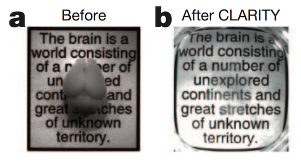
The opacity of the brain is one barrier to optically imaging individual neurons and their connections. Scattering in tissue is the main reason tissue is not transparent; absorption also plays a role…


Commoditizing population calcium imaging analysis

More open source software to check out.

Two-Photon Processor and SeNeCA – A freely available software package…


HelioScan software for imaging

HelioScan is a LabVIEW-based software suite from Fritjof Helmchen’s group that is specialized for running 2 photon microscopes, and is carefully designed to be modular. This modularity is intended to make it more easily…


Scientifica’s 2p scope

With its newly introduced scan head, Scientifica’s 2p scope is finally a complete package. They’re happy to sell their modular design in pieces, so this fills the void between fully custom rigs and turnkey…


DIY Superresolution, part II

This paper (York et al. 2012) came out earlier this year, but I thought it’s worth highlighting here, give the subject matter.

We present a hybrid technique, multifocal SIM (MSIM), that combines the resolution-doubling characteristics…


Micro-Manager

Micro-Manager (µ-Manager) is an open, ImageJ-based suite of code for controlling microscopes and associated instrumentation. Check out the list of supported equipment.


Whole embryo imaging

Drosophila embryos are a fraction of a square mm, and go from fertilization to hatching (as a larva) in about 22 hrs. So it’s possible to image individual embryos in their entirety…


Microbehunter

Oliver Kim puts out a nice magazine called Microbehunter. It’s a great resource for microscope nerds. I’ve listed a few blog articles below, but actually, the full PDF issues are where most of the…


Axial resolution and numerical aperture, part II

To recap the previous post on axial resolution and numerical aperture in two-photon microscopy:

For excitation deep in scattering tissue, higher NA can actually be detrimental because the light cone at…


Axial resolution vs numerical aperture

Recently, microscope manufacturers have been releasing ever higher NA objectives for multiphoton imaging. Although higher NA objectives should give better axial resolution, they might not be ideal for imaging deep into the…


Tsien’s VoltageFluors

Roger Tsien’s lab recently published the new generation voltage sensitive dye they were presenting at SfN: VoltageFluors. As often when then Tsien Lab takes on a new field, they start by taking a completely…


Fast z-galvo for 3D two-photon imaging

Perhaps you’re familiar with the various AOD and piezo-based methods for two-photon imaging in 3D quickly. Here’s another way (Botcherby et al. 2012, PNAS, open access)

The used all galvos, rather than AODs or…


Intrinsic imaging on the cheap

Intrinsic signal optical imaging is a functional imaging modality where the reflectance of red light indicates active portions of cortex. It is used for many applications, including imaging individual barrels in rodent somatosensory cortex, maps in visual cortex, and the tonotopic organization in auditory cortex.

Here is a prior Labrigger post with tips for intrinsic…


Clearing tissue for imaging: index matching is not new

The main factor that limits how deep we can image into tissue is the scattering of light. Multiphoton imaging partially mitigates the problem by using infrared light, which scatter less, and by using an excitation…


Open Microscopy Environment, for managing microscope images

The Open Microscopy Environment (OME) is a group of programmers, from academia and industry, who put together open source software packages for managing microscopy data. The emphasis is on handing multiple image formats, organizing…