Laser power modulation: EOMs and AOMs
Posted in Hardware
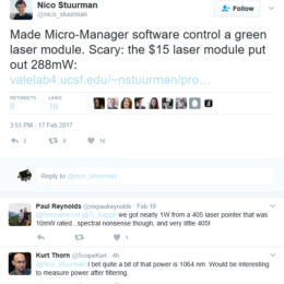
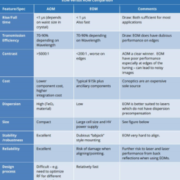
Fast laser power modulation in 2p systems is typically done with a Pockels cell. Conoptics sells one that is optimized for common Ti:Sapph systems, and that’s what many people end up using.
Pockels cells are a…